 interior of the cochlea (JPEG:70k).
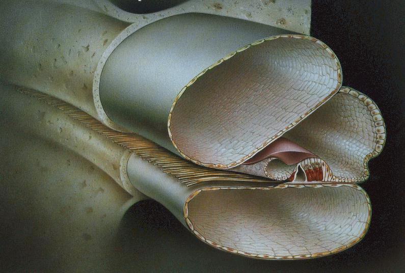
 Acoustic signals that enter the fluid-filled cochlear chambers, propagate in a dispersive manner along the cochlear partition. The partition, which spans the length and width of the cochlea, consists of the basilar membrane, tectorial membrane, and organ of Corti. The figure below shows an interior view of the cochlea.
interior of the cochlea (JPEG:70k).
The organ of Corti is a collection of cells, including the sensory hair cells, that sit on the basilar membrane. Along the upper surface of the organ of Corti (called the reticular lamina, RL) hair bundles (HB) protrude from the tops (or apexes) of the hair cells. Each bundle is composed of two to four rows of hair-like structures called stereocilia. Connected to the bottom (or base) of each hair cell are nerve fibers from the auditory nerve. There are two types of hair cells in the cochlea. The inner hair cells (IHC) are primarily innervated (that is, connected to the auditory nerve) by afferent fibers, which deliver neural signals to the brain. The outer hair cells (OHC), on the other hand, are innervated primarily by efferent nerve fibers, which receive neural signals from the brain.
 Organ of Corti
The human cochlea is believed to contain approximately 4000 IHCs and 12 000 OHCs, with four cells radially abreast and spaced every 10 microns along the basilar membrane's length. The tectorial membrane lies on top of the organ of Corti and is attached, at its inner edge, to the bony spiral limbus. A thin fluid space (4 to 6 microns) lies between these two surfaces, which shear as the basilar membrane moves up and down.
Hair cells are primarily mechano-electric transducers that convert displacement of the hair bundle (due to shearing between the tectorial membrane and the reticular lamina) into a change in the receptor current flowing through the cell. This is done by mechanical gating of ion channels that must be located in the hair bundle, probably near the top of each stereocilium. OHCs also act as electromechanic transducers by converting voltage across their cell membrane into length changes. This capability is important because it possible for the OHCs to influence cochlear micromechanics.
Each point on the basilar membrane is tuned to a different frequency, with a spatial gradient of about 0.2 octaves/mm for human, and about 0.32 octaves/mm for cat. Roughly speaking, the cochlea acts like a bank of filters. The filtering allows the separation of various frequency components of the signal with a good signal-to-noise ratio. However, each filter has dynamic range compression built into its mechanical response. This nonlinearity makes the frequency response of each filter dependent on the level of the acoustic signal.
All mammalian cochleas appear to function according to the same basic principles; however, the effective frequency range differs among species. For example, the range of audible frequencies is about 20 Hz to 16 kHz in the human cochlea and about 100 Hz to 40kHz in the cat cochlea. The human basilar membrane is about 35 mm long, while the cat basilar membrane is about 25 mm.
Return to Cochlear Mechanics or proceed to Physiological Measures.